
-
Biceps Tendon Tear at the Elbow

A biceps tear can be complete or partial. Partial biceps tendon tears will not completely break the tendon while complete tendon tears will break the tendon into two parts. Tears of the distal biceps tendon are usually complete and the muscle is separated from the bone. Tears of the distal biceps tendon most often result from a sudden injury or lifting a heavy object.
-
Elbow (Olecranon) Bursitis
Inflammation of the olecranon bursa leads to a condition called olecranon bursitis. The causes of elbow bursitis may include trauma or a hard blow on the elbow, excessive leaning on the elbow, infection by puncture wounds or insect bites, or conditions such as gout and rheumatoid arthritis.
-
Elbow Dislocation
The arm in the human body is made up of three bones that join to form a hinge joint called the elbow. The upper arm bone or humerus connects from the shoulder to the elbow to form the top of the hinge joint. The lower arm or forearm consists of two bones, the radius, and the ulna. These bones connect the wrist to the elbow forming the bottom portion of the hinge joint.
-
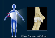
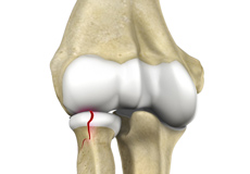
Elbow Fractures in Children
Children’s bones have an area of developing cartilage tissue called a growth plate present at the end of long bones that will eventually develop into solid bone as the child grows. Fractures are more common in children due to their physical activities as well as their bone properties. An elbow fracture most commonly occurs when your child falls on an outstretched arm.
-
Radial Head Fractures
Radial head fractures are very common and occur in almost 20% of acute elbow injuries. Elbow dislocations are generally associated with radial head fractures. Radial head fractures are more common in women than in men and occur more frequently in the age group of 30 to 40 years.
-
Tennis Elbow
Tennis elbow is a common name for the elbow condition lateral epicondylitis. It is an overuse injury that causes inflammation and microtears of the tendons that attach to the lateral epicondyle.
-
Throwing Injuries in the Elbow
An athlete uses an overhand throw to achieve greater speed and distance. Repeated throwing in sports such as baseball and basketball can place a lot of stress on the joints of the arm, and lead to weakening and ultimately, injury to the structures in the elbow.
-
Ulnar Nerve Entrapment
When the elbow is bent, the ulnar nerve can stretch and catch on the bony bump. When the ulnar nerve is compressed or entrapped, the nerve can tear and become inflamed, leading to cubital tunnel syndrome.


Ulnar Nerve Release

Ulnar nerve release, also known as ulnar nerve decompression, is a surgical procedure to treat a medical condition called ulnar nerve entrapment.
Elbow Surgery

Elbow surgery is a surgical procedure for the treatment of an elbow injury or elbow condition. The procedure involves repairing a diseased, damaged, or degenerated elbow joint in order to eliminate pain and restore normal function.
Elbow Arthroscopy

Elbow arthroscopy, also referred to as keyhole or minimally invasive surgery, is a surgical procedure that is performed through tiny incisions to evaluate and treat several elbow conditions.
The elbow is a complex joint formed by the articulation of three bones – the humerus, radius, and ulna. The elbow joint helps in bending or straightening of the arm to 180 degrees and lifting or moving objects.
The bones of the elbow are supported by:
- Ligaments and tendons
- Muscles
- Nerves
- Blood vessels
Bones and Joints of the Elbow
The elbow joint is formed at the junction of three bones:
- The humerus (upper arm bone) forms the upper portion of the joint. The lower end of the humerus divides into two bony protrusions known as the medial and lateral epicondyles, which can be felt on either side of the elbow joint.
- The ulna is the larger bone of the forearm located on the inner surface of the joint. It articulates with the humerus.
- The radius is the smaller bone of the forearm situated on the outer surface of the joint. The head of the radius is circular and hollow, which allows movement with the humerus. The articulation between the ulna and radius helps the forearm to rotate.
The elbow consists of three joints, namely:
- The humeroulnar joint is formed between the humerus and ulna and allows flexion and extension of the arm.
- The humeroradial joint is formed between the radius and humerus and allows movements like flexion, extension, supination, and pronation.
- The radioulnar joint is formed between the ulna and radius bones and allows rotation of the lower arm.
Articular cartilage lines the articulating regions of the humerus, radius, and ulna. It is a thin, tough, flexible and slippery surface that acts as a shock absorber and cushion to reduce friction between the bones. The cartilage is lubricated with synovial fluid, which further enables the smooth movement of the bones.
Muscles of the Elbow Joint
There are several muscles extending across the elbow joint that help in various movements. These include the following:
- Biceps brachii: Upper arm muscle, enabling flexion of the arm
- Triceps brachii: Muscle in the back of the upper arm that extends the arm and fixes the elbow during fine movements
- Brachialis: Upper arm muscle beneath the biceps, which flexes the elbow towards the body
- Brachioradialis: Forearm muscle that flexes, straightens and pulls the arm at the elbow
- Pronator teres: Muscle that extends from the humeral head, across the elbow, and towards the ulna, and helps to turn the palm facing backward
- Extensor carpi radialis brevis: Forearm muscle that helps in movement of the hand
- Extensor digitorum: Forearm muscle that helps in movement of the fingers
Ligaments and Tendons of the Elbow
The elbow joint is supported by ligaments and tendons, which provide stability to the joint.
Ligaments are a group of firm tissues that connect bones to other bones. The most important ligaments of the elbow joint are the:
- Medial or ulnar collateral ligament: Comprised of triangular bands of tissue on the inner side of the elbow joint
- Lateral or radial collateral ligament: A thin band of tissue on the outer side of the elbow joint
- Annular ligament: Group of fibers that surround the radial head, and hold the ulna and radius tightly in place during movement of the arm
Together, the medial and lateral ligaments are the main source of stability and hold the humerus and ulna tightly in place during movement of the arm.
The ligaments around a joint combine to form a joint capsule that contains synovial fluid.
Any injury to these ligaments can lead to instability of the elbow joint.
Tendons are bands of connective tissue fibers that connect muscle to bone. The various tendons that surround the elbow joint include:
- Biceps tendon: attaches the biceps muscle to the radius, allowing the elbow to bend
- Triceps tendon: attaches the triceps muscle to the ulna, allowing the elbow to straighten
Nerves of the Elbow
The main nerves of the elbow joint are the ulnar, radial and median nerves. These nerves transfer signals from the brain to the muscles that aid in elbow movements. They also carry sensory signals such as touch, pain, and temperature back to the brain.
Any injury or damage to these nerves causes pain, weakness or joint instability.
Blood Vessels Supplying the Elbow
Arteries are blood vessels that carry oxygen-pure blood from the heart to the hand. The main artery of the elbow is the brachial artery that travels across the inside of the elbow and divides into two small branches below the elbow to form the ulnar and the radial artery.